Physeal-Sparing MPFL Reconstruction in Adolescents
An Interview with:
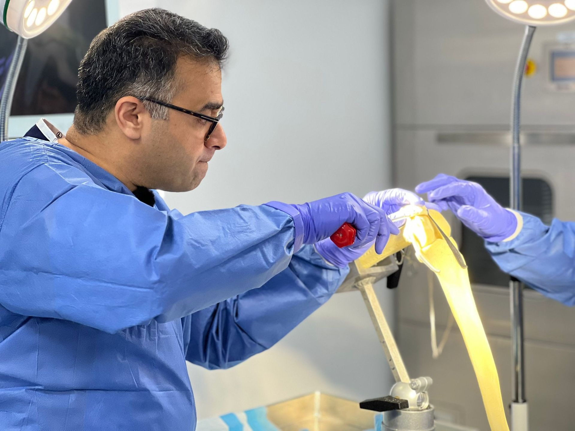
Dr. Nayef Aslam-Pervez
Issue 20, April 2026

What led you to develop a physeal- sparing approach for MPFL reconstruction in younger patients? Was there a particular case or clinical gap that pushed you toward it?

There wasn’t a single defining case, but
rather a progressive shift in my clinical
practice. I became the primary referral
surgeon for paediatric patellofemoral
instability within my institution, managing
approximately 50 such cases per year.
With that volume, it became increasingly
important to optimise outcomes in a group
of patients who present unique anatomical
and developmental considerations.
Attending specialist meetings, including
paediatric knee conferences such as the Kid
Knee meeting in Sheffield, further
highlighted the need to refine our approach.
This led me to focus on developing a
technique that addresses instability in an
anatomical and reproducible way, while
respecting the distal femoral physis and
avoiding unnecessary donor site morbidity
by preserving the hamstrings.
In patients who are approaching skeletal maturity, what are the main technical or decision-making challenges when planning MPFL reconstruction?
The main challenge lies in the variability of
skeletal maturity in this age group.
Chronological age does not always reflect
skeletal maturity, and surgeons must carefully
assess the relationship between the planned
femoral fixation point and the distal femoral
physis. Another important consideration is
balancing stability with the risk of over-
constraint. Adolescents may still undergo some
degree of growth, so reconstruction must
restore stability without excessively tightening
the medial restraint of the patellofemoral joint.
This becomes particularly relevant when using
synthetic ligaments. At this stage of
development, growth is predominantly
longitudinal rather than radial, and overall knee
dimensions are approaching adult morphology.
As a result, the risk of progressive over-
tensioning of a non-elastic synthetic MPFL
construct is likely reduced compared to
younger children.
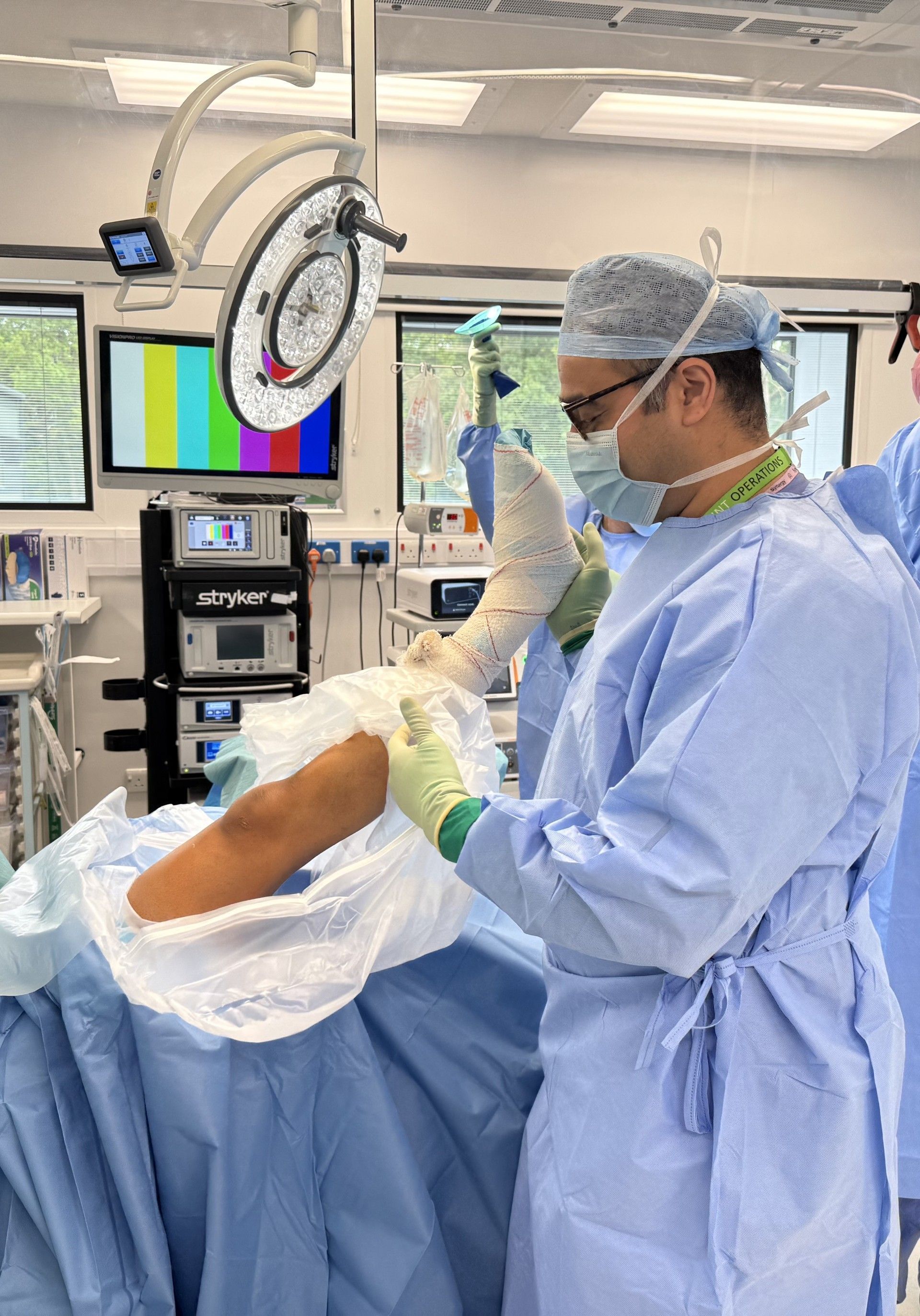
Can you walk us through how you use intraoperative fluoroscopy to protect the distal femoral physis, and what surgeons should be looking for in real time?
Fluoroscopy plays a critical role in safely
identifying the femoral insertion point
while protecting the distal femoral physis.
Research has highlighted that Schöttle’s
point is consistently located distal to the
medial femoral physis, which aligns with
my clinical experience.
Intraoperatively, I begin by obtaining a
true lateral radiograph to identify
Schöttle’s point. On this view, the entry
point may appear to overlap or even
traverse the physis. However, this can be
misleading due to the characteristic “W-
shaped” morphology of the distal femoral
physis. The medial limb of this “W” slopes
proximally, creating the illusion that the
entry point lies within the physis on the
lateral projection.
For this reason, I always confirm
positioning with an AP view before
entering the bone. The AP radiograph
reliably demonstrates that Schöttle’s
point is distal to the physis on the medial
side. Once confirmed, the Beath pin is
placed onto the periosteum and gently
tapped into the epiphysis rather than
drilled, minimising the risk of thermal
injury.
The guidewire is directed slightly
anteriorly and distally, ensuring that the
femoral socket remains entirely within
the epiphysis and away from the physis. A
hand reaming technique is then used to
create the socket, further reducing the
risk of thermal damage.
Although some adult techniques rely on
anatomical landmarks without
fluoroscopy, I would strongly advocate
routine use of image intensification in
paediatric cases, where direct
visualisation of the physis is essential to
ensure safe and accurate tunnel
placement.
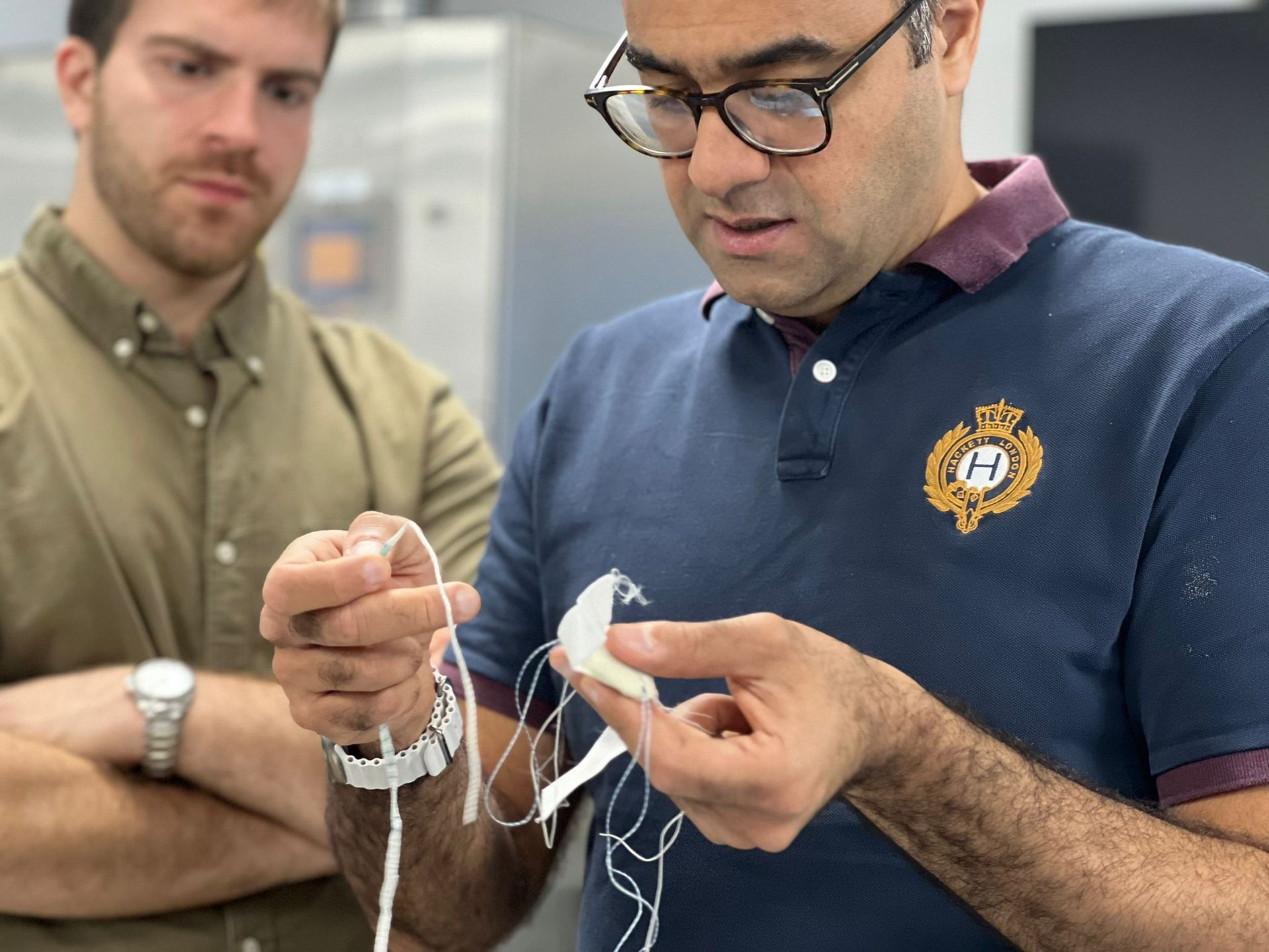
You’ve chosen a synthetic ligament for this technique. What was the rationale behind that choice, and what advantages do you feel it offers compared to autograft or allograft in this age group?
In adolescent patients, avoiding donor-site
morbidity is an important consideration.
Harvesting hamstring tendons can lead to
measurable strength deficits, and in younger
patients the gracilis tendon is often relatively
small, occasionally necessitating harvest of the
semitendinosus to achieve an adequate graft.
Preserving these structures is particularly
valuable in active patients.
In addition, many patients presenting with
patellofemoral instability in this age group
demonstrate features of generalized
ligamentous laxity. In such cases, autograft
tissue may itself be relatively compliant, raising
the possibility of graft elongation over time. A
synthetic ligament offers a more consistent
construct, which may be advantageous in this
cohort.
There is also emerging clinical evidence
supporting the use of synthetic ligaments in
MPFL reconstruction. Work by Hersh Deo using
the XIROS 5mm Infinity Lock tape has
demonstrated excellent clinical outcomes with
low failure rates, providing a strong foundation
for further application in selected patients.
From a technical perspective, the XIROS Infinity
Loop tape has a low-profile, flat configuration,
which is well suited to the relatively superficial
anatomy of the MPFL, particularly in paediatric
patients. Its open-weave structure also allows
for tissue ingrowth and biological integration
over time.
In my early experience, patients have
demonstrated a rapid recovery and high levels
of satisfaction. While there is a theoretical risk of
over-constraint with non-elastic constructs,
careful patient selection and appropriate
intraoperative tensioning are key. We are
currently undertaking structured follow-up of
our paediatric cohort and hope to report these
outcomes in due course.
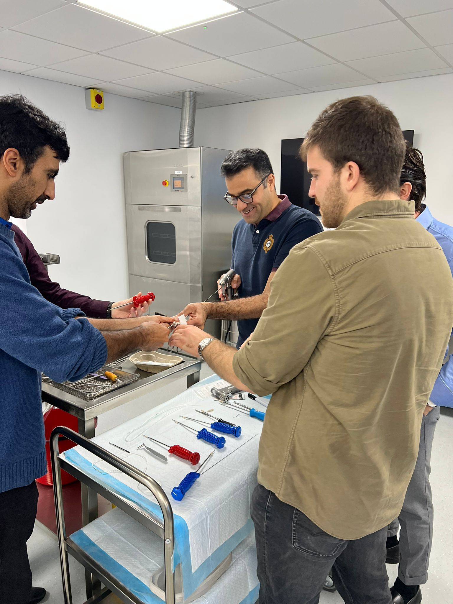
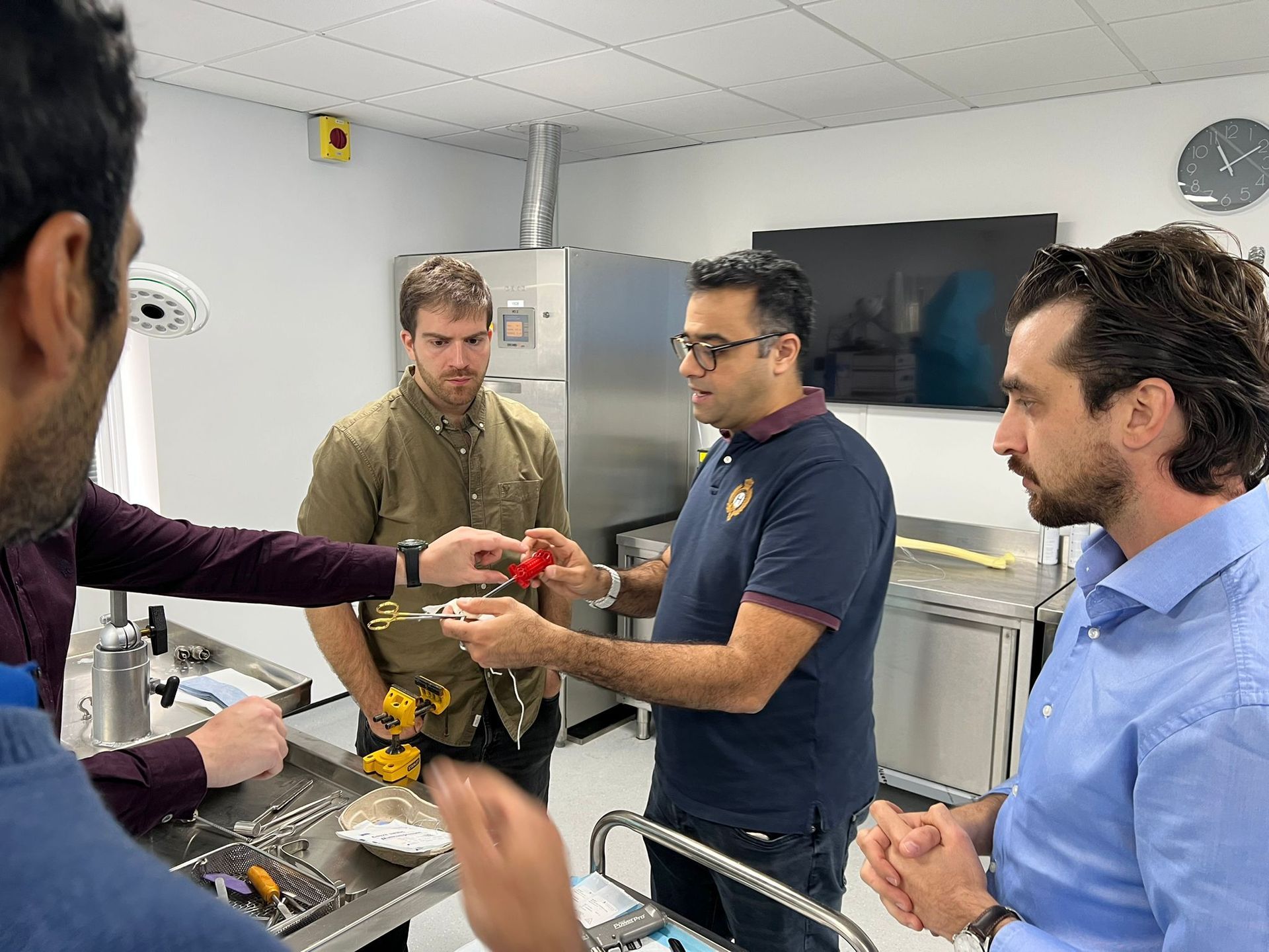
What are the key technical points surgeons need to get right, particularly around patellar preparation and femoral fixation, to minimise complications?
One of the key differences in this
technique is that it reverses the traditional
sequence of fixation. Rather than fixing on
the patella first and tensioning on the
femoral side, I perform femoral fixation
initially using a blind-ending socket within
the distal femoral epiphysis, and then
tension the construct on the patellar side.
This approach allows much more
controlled assessment of patellar tracking.
By passing the synthetic tape through
two transverse patellar tunnels and
temporarily securing it on the lateral side
with a clamp, I can assess patellar stability
dynamically through a full range of
motion. This includes full extension, 30
degrees, and 60 degrees of flexion, while
also evaluating lateral translation. It allows
both hands to be free, facilitating a more
accurate and reproducible assessment of
appropriate tensioning.
On the patellar side, careful preparation is
essential to minimise the risk of fracture.
The use of small-diameter (2.4 mm)
transverse tunnels creates a minimal bony
footprint and reduces stress risers. Once
appropriate tension is confirmed, final
fixation is performed on the medial side
using PEEK bone anchors, typically 3.5
mm or 4.75 mm depending on patellar
size, which can be planned preoperatively
using imaging. Importantly, only the
medial portion of the patella requires
preparation for anchor placement, rather
than full-length drilling across the entire
patella, further reducing risk.
Additional technical considerations
include the use of a tap in patients with
denser bone to facilitate anchor insertion
and minimise stress on the patella. Early
in the learning curve, an ACL guide can
assist with accurate tunnel placement,
although this becomes less necessary
with experience. Overall, careful attention to tunnel size,
controlled tensioning, and dynamic
assessment of patellar tracking are key to
achieving a stable reconstruction while
avoiding complications such as patellar
fracture or over-constraint.
At what degree of flexion are you fixing the graft, and how do you assess appropriate tension in these younger patients?
The graft is typically fixed with the knee in
approximately 30 degrees of flexion. At this
position, the patella is beginning to engage the trochlea, allowing a reliable assessment of medial restraint. In this technique, I tension the graft on the
patellar side rather than the femoral side. By
temporarily securing the synthetic tape on the lateral side of the patella with a clamp, I am able to assess patellar tracking dynamically using both hands. This allows evaluation of lateral translation and tracking through a range of motion,
including full extension, 30 degrees, and 60
degrees of flexion. In my experience, this provides a more controlled and reproducible method of assessing tension compared to femoral-side tensioning, where
manual control can be more limited.
It is important that the graft functions as a check- rein rather than a rigid restraint. In particular,
when using a synthetic ligament in this age
group, it is preferable to err slightly on the side of a looser construct rather than risking over-constraint. Excessive tension should be avoided to minimise the risk of medial patellofemoral overload and altered joint mechanics.
From your early cases, what have you observed in terms of stability, return to activity, and complication rates?
In our early experience, patients have
demonstrated excellent restoration of
patellar stability following synthetic MPFL
reconstruction. Recovery has been relatively
rapid, which we attribute in part to avoiding
hamstring harvest and therefore reducing
overall surgical morbidity.
Patients have reported high levels of
satisfaction and have been able to return to
their previous activities, and in some cases
exceed their pre-injury level of function.
Importantly, we have not observed any
cases of over-constraint with the synthetic
construct when appropriate tensioning
principles are followed. Radiographic
follow-up with long-leg alignment films has
not demonstrated any change in coronal
alignment, and we have not identified any
evidence of physeal injury when femoral
fixation is performed using a careful,
fluoroscopy-guided technique.
We have also performed postoperative MRI
assessments in our cohort, which have not
demonstrated any evidence of patellar tilt
or medial over-constraint. These imaging
findings provide further reassurance that
the reconstruction restores stability without
adversely affecting patellofemoral
mechanics.
Our early cohort has now been followed for
at least one year, and complication rates to
date have been very low. We are continuing
structured follow-up, including comparison
of pre- and postoperative MRI findings, to
better understand how the synthetic
ligament contributes to stability over time
and how it behaves in relation to femoral
growth as patients continue to develop. We
hope to present and publish these results
to further inform clinical practice.
How do you see MPFL reconstruction evolving in paediatric and adolescent patients over the next five to ten years, and what role do you think synthetic reconstruction will play?
There is an increasing recognition that
patellofemoral instability is multifactorial, and not solely the result of MPFL insufficiency. Factors such as trochlear morphology, coronal and rotational alignment, and generalized
ligamentous laxity all play an important role, and need to be considered when planning treatment. As a result, the future of MPFL reconstruction in paediatric and adolescent patients is likely to
move toward a more individualised, anatomy-driven approach, where reconstruction is combined with correction of underlying risk factors when necessary.
In addition, there is growing interest in
alternative medial stabilising structures such as the medial quadriceps tendon–femoral ligament (MQTFL), which attaches from the distal quadriceps tendon to the femur near Schöttle’s point. This may offer another option for restoring medial restraint, particularly in patients where
patellar fixation is less desirable.
We are also likely to see increasing comparative data on different graft choices, including hamstring autograft, quadriceps-based techniques, and synthetic ligaments. As this evidence evolves, it will allow for more tailored, patient-specific surgical decision-making. Over the next five to ten years, we can expect more robust long-term outcome data to emerge, not only for isolated MPFL reconstruction but also for combined procedures addressing these
contributing factors. This will allow us to better understand which patients benefit from additional interventions and how best to optimise long-term joint health.
From a clinical perspective, we are already seeing the long-term consequences of untreated instability, with patients presenting later in life with patellofemoral degeneration requiring joint replacement. Earlier and more comprehensive management of instability may help to preserve joint function and delay or prevent this progression. Synthetic reconstruction is likely to play an increasing role in selected patients, particularly where preservation of native tissues is desirable. As further outcome data becomes available, its role will become more clearly defined within the broader management strategy for
patellofemoral instability.
About Dr. Nayef Aslam-Pervez
Mr. Aslam-Pervez is a distinguished Consultant Orthopaedic Surgeon based in Yorkshire. He works at a major trauma centre and tertiary referral hospital, treating highly complex cases
with excellent outcomes.
The Knee Research Unit (KRUH) at Hull
University Teaching Hospitals was established to lead and coordinate knee-related research within the Trust and as part of a regional MDT across Yorkshire. The unit is involved in multiple research projects, including the paediatric synthetic MPFL reconstruction study, with ongoing follow-up and further research being conducted under the umbrella
of the KRUH.
As the Lead for Soft Tissue Knee Surgery and previously Clinical Governance Lead in both Hull and London, Mr. Aslam-Pervez has extensive experience in building and
maintaining the highest quality of orthopaedic services. His practice combines advanced surgical
techniques with comprehensive patient care to deliver excellent outcomes, with a special interest in sports injuries and joint replacement surgery.
We would like to thank Dr. Nayef Aslam-Pervez for his insight.
Thank you for being part of Our Community
We believe in pushing boundaries, sharing knowledge, and highlighting the people and ideas shaping the future of orthopaedics. On the Podium is our platform for showcasing the journeys, challenges, and breakthroughs of those making a positive impact in the orthopaedic community.
If you’ve found value in these stories, we encourage you to stay connected. Follow us on social media for the latest updates, subscribe to our email list for exclusive content, and share these insights with others who might find them just as inspiring.
Your engagement helps us continue to bring important stories to light and grow a community of forward thinkers.
Thank you for reading, supporting, and being part of this journey. We look forward to exploring more with you.
Thank you to our sponsors:


To download this issue of On The Podium, click below.
Sign up to On The Podium. The latest insights and inspiration from orthopaedic specialists around the globe sent straight to your inbox.
On The Podium
The newsletter brought to you by OrthoSpaceX
| Engage with Orthopaedic Leaders | Exclusive orthopaedic insight | |
| Stay Ahead in Orthopaedic Advancements | Uncover Surgical Innovations | |
| Empower Your Orthopaedic Knowledge | Unlock the Minds of Orthopaedic Specialists |